HABs taxon details
Alexandrium tamarense (Lebour) Balech, 1995
109714 (urn:lsid:marinespecies.org:taxname:109714)
accepted
Species
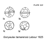
Gonyaulax tamarensis Lebour, 1925 · unaccepted (basionym)
marine
(of Gonyaulax tamarensis Lebour, 1925) Lebour, M.V. (1925). The Dinoflagellates of Northern Seas. <em>The Marine Biological Association of the United Kingdom.</em> 250 pp. [details] Available for editors 
Type locality contained in Tamar estuary
type locality contained in Tamar estuary [details]
Harmful effect For a discussion of the species in the tamarense group, see under A. catenella. John et al. (2014) have suggested that the...
Identification This species, belonging to the Alexandrium tamarense species complex, can only be unambiguously identified using genetic...
Verified sequences Strain CCMP115 (Higman et al. 2001; Orr et al. 2011; Zou et al. 2014):
SSU/ITS/5.8S/ITS2/LSU rDNA JF521640
LSU rDNA AJ308587
Harmful effect For a discussion of the species in the tamarense group, see under A. catenella. John et al. (2014) have suggested that the name A. tamarense be applied to isolates belonging to Group II. With this concept A. tamarense would be a harmless species.
Only a strain (CNR-ATA6PT) from Sardinia was reported to have GTX (Gonyautoxins) once analysed by high pressure liquid chromatography coupled to a mass spectrometry (HPLC/MS) (Lugliè et al. 2017). [details]
Only a strain (CNR-ATA6PT) from Sardinia was reported to have GTX (Gonyautoxins) once analysed by high pressure liquid chromatography coupled to a mass spectrometry (HPLC/MS) (Lugliè et al. 2017). [details]
Identification This species, belonging to the Alexandrium tamarense species complex, can only be unambiguously identified using genetic...
Identification This species, belonging to the Alexandrium tamarense species complex, can only be unambiguously identified using genetic techniques. [details]
Verified sequences Strain CCMP115 (Higman et al. 2001; Orr et al. 2011; Zou et al. 2014):
SSU/ITS/5.8S/ITS2/LSU rDNA JF521640
LSU rDNA AJ308587
Verified sequences Strain CCMP115 (Higman et al. 2001; Orr et al. 2011; Zou et al. 2014):
SSU/ITS/5.8S/ITS2/LSU rDNA JF521640
LSU rDNA AJ308587
5.8S/ITS2 rDNA HM483847 [details]
SSU/ITS/5.8S/ITS2/LSU rDNA JF521640
LSU rDNA AJ308587
5.8S/ITS2 rDNA HM483847 [details]
Guiry, M.D. & Guiry, G.M. (2025). AlgaeBase. World-wide electronic publication, National University of Ireland, Galway (taxonomic information republished from AlgaeBase with permission of M.D. Guiry). Alexandrium tamarense (Lebour) Balech, 1995. Accessed through: Lundholm, N.; Bernard, C.; Churro, C.; Escalera, L.; Hoppenrath, M.; Iwataki, M.; Larsen, J.; Mertens, K.; Moestrup, Ø.; Murray, S.; Salas, R.; Tillmann, U.; Zingone, A. (Eds) (2009 onwards) IOC-UNESCO Taxonomic Reference List of Harmful Micro Algae at: https://www.marinespecies.org/hab/aphia.php?p=taxdetails&id=109714 on 2025-07-10
Lundholm, N.; Bernard, C.; Churro, C.; Escalera, L.; Hoppenrath, M.; Iwataki, M.; Larsen, J.; Mertens, K.; Moestrup, Ø.; Murray, S.; Salas, R.; Tillmann, U.; Zingone, A. (Eds) (2009 onwards). IOC-UNESCO Taxonomic Reference List of Harmful Micro Algae. Alexandrium tamarense (Lebour) Balech, 1995. Accessed at: https://www.marinespecies.org/hab/aphia.php?p=taxdetails&id=109714 on 2025-07-10
Date
action
by
2006-07-27 06:59:07Z
changed
Camba Reu, Cibran
2015-06-26 12:00:51Z
changed
db_admin
original description
(of Gonyaulax tamarensis Lebour, 1925) Lebour, M.V. (1925). The Dinoflagellates of Northern Seas. <em>The Marine Biological Association of the United Kingdom.</em> 250 pp. [details] Available for editors 
basis of record Guiry, M.D. & Guiry, G.M. (2025). AlgaeBase. <em>World-wide electronic publication, National University of Ireland, Galway.</em> searched on YYYY-MM-DD., available online at http://www.algaebase.org [details]
basis of record Gómez, F. (2005). A list of free-living dinoflagellate species in the world's oceans. <em>Acta Bot. Croat.</em> 64(1): 129-212. [details]
additional source Orr, R. J.; Stüken, A.; Rundberget, T.; Eikrem, W.; Jakobsen, K. S. (2011). Improved phylogenetic resolution of toxic and non-toxic <i>Alexandrium</i> strains using a concatenated rDNA approach. <em>Harmful Algae.</em> 10(6): 676-688., available online at https://doi.org/10.1016/j.hal.2011.05.003 [details]
additional source Balech, E. (2002). Dinoflagelados tecados tóxicos en el Cono Sur Americano. <em>In: Sar, E.A., Ferrario, M.E. & Reguera, B. (Eds.). Floraciones Algales Nocivas en el Cono Sur Americano. Instituto Español de Oceanografía.</em> pp. 123-144. [details] Available for editors
additional source Higman, W. A.; Stone, D. M.; Lewis, J. M. (2001). Sequence comparisons of toxic and non-toxic <i>Alexandrium tamarense</i> (Dinophyceae) isolates from UK waters. <em>Phycologia.</em> 40(3): 256-262., available online at https://doi.org/10.2216/i0031-8884-40-3-256.1 [details]
additional source Zou, C.; Ye, R.-M.; Zheng, J.-W.; Luo, Z.-H.; Gu, H.-F.; Yang, W.-D.; Li, H.-Y.; Liu, J.-S. (2014). Molecular phylogeny and PSP toxin profile of the <i>Alexandrium tamarense</i> species complex along the coast of China. <em>Marine Pollution Bulletin.</em> 89(1-2): 209-219., available online at https://doi.org/10.1016/j.marpolbul.2014.09.056 [details]
additional source Guiry, M.D. & Guiry, G.M. (2025). AlgaeBase. <em>World-wide electronic publication, National University of Ireland, Galway.</em> searched on YYYY-MM-DD., available online at http://www.algaebase.org [details]
additional source Tomas, C.R. (Ed.). (1997). Identifying marine phytoplankton. Academic Press: San Diego, CA [etc.] (USA). ISBN 0-12-693018-X. XV, 858 pp., available online at http://www.sciencedirect.com/science/book/9780126930184 [details]
additional source Brandt, S. (2001). Dinoflagellates, <B><I>in</I></B>: Costello, M.J. <i>et al.</i> (Ed.) (2001). <i>European register of marine species: a check-list of the marine species in Europe and a bibliography of guides to their identification. Collection Patrimoines Naturels,</i> 50: pp. 47-53 (look up in IMIS) [details]
additional source Scott, F.J.; Marchant, H.J. (Ed.). (2005). Antarctic marine protists. <em>Australian Biological Resources Study: Canberra.</em> ISBN 0-642-56835-9. 563 pp., available online at http://its-db.aad.gov.au/proms/pubn/pubshow.asp?pub_id=12140 [details]
additional source Balech, E. 1995. The Genus <i>Alexandrium</i> Halim (Dinoflagellata). Sherkin Island Marine Station, Sherkin Island, Co. Cork, Ireland, 151. [details] Available for editors
additional source Moestrup, Ø., Akselman, R., Cronberg, G., Elbraechter, M., Fraga, S., Halim, Y., Hansen, G., Hoppenrath, M., Larsen, J., Lundholm, N., Nguyen, L. N., Zingone, A. (Eds) (2009 onwards). IOC-UNESCO Taxonomic Reference List of Harmful Micro Algae., available online at http://www.marinespecies.org/HAB [details]
additional source Chang, F.H.; Charleston, W.A.G.; McKenna, P.B.; Clowes, C.D.; Wilson, G.J.; Broady, P.A. (2012). Phylum Myzozoa: dinoflagellates, perkinsids, ellobiopsids, sporozoans, in: Gordon, D.P. (Ed.) (2012). New Zealand inventory of biodiversity: 3. Kingdoms Bacteria, Protozoa, Chromista, Plantae, Fungi. pp. 175-216. [details]
redescription John, U.; Litaker, R. W.; Montresor, M.; Murray, S.; Brosnahan, M. L.; Anderson, D. M. (2014). Formal revision of the <i>Alexandrium tamarense</i> species complex (Dinophyceae) taxonomy: The introduction of five species with emphasis on molecular-based (rDNA) cassification. <em>Protist.</em> 165(6): 779-804., available online at https://doi.org/10.1016/j.protis.2014.10.001 [details]
new combination reference Balech, E. (1985). The genus <i>Alexandrium</i> or <i>Gonyaulax</i> of the <i>tamarensis</i> group. In: D.M. Anderson, A.W. White and D.G. Baden (Eds.) Toxic Dinoflagellates. New York: Elsevier. P. 33–38. [details] Available for editors
toxicology source Lugliè, A.; Giacobbe, M. G.; Riccardi, E.; Bruno, M.; Pigozzi, S.; Mariani, M. A.; Satta, C. T.; Stacca, D.; Bazzoni, A. M.; Caddeo, T.; Farina, P.; Padedda, B. M.; Pulina, S.; Sechi, N.; Milandri, A. (2017). Paralytic Shellfish Toxins and Cyanotoxins in the Mediterranean: New Data from Sardinia and Sicily (Italy). <em>Microorganisms.</em> 5(4): 72., available online at https://doi.org/10.3390/microorganisms5040072
note: A. tamarense strain CNR–ATA6PT showed moderate toxicity, especially due to GTX5, GTX1 and GTX4 [details] Available for editors
ecology source Jeong, H.; Yoo, Y.; Park, J.; Song, J.; Kim, S.; Lee, S.; Kim, K.; Yih, W. (2005). Feeding by phototrophic red-tide dinoflagellates: five species newly revealed and six species previously known to be mixotrophic. <em>Aquatic Microbial Ecology.</em> 40: 133-150., available online at https://doi.org/10.3354/ame040133 [details]
 Present
  Inaccurate
  Introduced: alien
  Containing type locality
Unknown type CCMP115, geounit Tamar estuary [details]
From regional or thematic species database
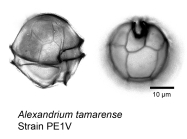
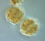
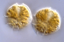
Description Cell is small- to relatively large-sized and is somewhat isodiametric. In ventral view, the shape is irregularly pentagonal and convex. The cell frequently has one or two shoulders that may or may not be very noticeable. The hypotheca is irregularly trapezoidal with convex and sometimes irregular sides. A concavity may be located on the left side of the hypotheca; sometimes a protuberance is above it. This concavity is not deep, but it is noticeable. The posterior margin is very often asymmetric, sloped forwards and to the right. The descending (1) cingulum is excavated and has a very narrow list. The sulcus is variably deep and has moderately developed lists. The PO is often very wide and markedly angular. Its dorsal margin is relatively extensive and is more often straight or nearly straight, but it is sometimes regular or irregularly convex. Commonly, the right margin is composed of two segments. The dorsal segment is usually straight or almost straight but is sometimes regularly or irregularly convex and parallel to the major axis of the plate. The ventral segment is sloped inward and is straight or rather concave. The ventral end is almost always fully truncated and forms a clear connection with 1'. Rarely, a PO is found that has a laterally located connecting pore that is almost always small (several were found in material from Norway [Oslo]). Small marginal pores were detected in the PO of thecae from England (Cornwall) and Korea. The 1' has a relatively variable width. Usually, the anterior angle and, especially, the posterior angle are rather extensively truncated. The anterior right margin is often rather concave. Sometimes, the margin is abruptly angled at the location of the ventral pore. Occasionally, the two larger margins are straight. The ventral pore is small and always exists. Both its position on. the margin and the degree of its indentation on the plate may vary. For example, the notch frequently occurs mostly within the suture region and penetrates only a little or not at all onto the plate. In other cases, the pore may be formed by two conjugated notches, one in 1' and the other in 4'. In some individuals, the notch is in the 4' plate, completely or almost completely. In this latter circumstance, the pore would be missed if the observer examined only the 1 '. 3' is always clearly asymmetrical, but the degree of asymmetry varies. 1 """" is somewhat narrow. The most variable feature is the development of the left sulcal list, which is rather wide in some cases and rather narrow in others. The right sulcal list, supported by the 5""' plate, is usually barely conspicuous. 2"""" is variable. It is transversely extended in some thecae (type B) or dorsoventrally in others (type A), with some in transition. Type B is dominant and is the type represented in Lebour's description. However, the transition forms occur frequently. The sulcal plates also vary. The S.a. is always narrow, is generally a little longer than wide, and has a deep posterior sinus. The anterior margin is more or less straight, wide, and situated at or a little above the level of the anterior left margin of the cingulum. In Lebour's figure, the anterior margin clearly intrudes onto the epitheca. From the left side of this margin, a short and oblique groove is directed to the right and down. Frequently, a portion of this margin, usually the right part but sometimes the left part, is lifted more or less abruptly and penetrates into the epitheca, sometimes quite deeply. This may be a kequent and rather noticeable characteristic in cultured specimens, but it is rare in field specimens. The S.p. may or may not have a connecting pore that is small, clearly displaced to the right, and connected to the edge by a groove. Sometimes, a groove occurs without a pore; many thecae have neither, as is the case for most of those from the Argentine Sea. The length/width relation of this plate is somewhat variable. Of the remaining sulcal plates, the most variable [details]Harmful effect For a discussion of the species in the tamarense group, see under A. catenella. John et al. (2014) have suggested that the name A. tamarense be applied to isolates belonging to Group II. With this concept A. tamarense would be a harmless species.
Only a strain (CNR-ATA6PT) from Sardinia was reported to have GTX (Gonyautoxins) once analysed by high pressure liquid chromatography coupled to a mass spectrometry (HPLC/MS) (Lugliè et al. 2017). [details]
Identification This species, belonging to the Alexandrium tamarense species complex, can only be unambiguously identified using genetic techniques. [details]
Verified sequences Strain CCMP115 (Higman et al. 2001; Orr et al. 2011; Zou et al. 2014):
SSU/ITS/5.8S/ITS2/LSU rDNA JF521640
LSU rDNA AJ308587
5.8S/ITS2 rDNA HM483847 [details]
Published in AlgaeBase 
Published in AlgaeBase (from synonym Gonyaulax tamarensis Lebour, 1925)
Published in AlgaeBase (from synonym Gessnerium tamarensis (Lebour) Loeblich III & L.Loeblich, 1979)
To Barcode of Life (6 barcodes)
To Biodiversity Heritage Library (10 publications)
To Biodiversity Heritage Library (2 publications) (from synonym Gessnerium tamarensis (Lebour) Loeblich III & L.Loeblich, 1979)
To Biodiversity Heritage Library (23 publications) (from synonym Gonyaulax tamarensis Lebour, 1925)
To Dyntaxa
To European Nucleotide Archive, ENA (Alexandrium tamarense)
To GenBank (683291 nucleotides; 144 proteins)
To GenBank (683291 nucleotides; 144 proteins) (from synonym Gonyaulax tamarensis Lebour, 1925)
To Global Biotic Interactions (GloBI)
To Information system on Aquatic Non-Indigenous and Cryptogenic Species (AquaNIS)
To PESI
To PESI (from synonym Gonyaulax tamarensis Lebour, 1925)
To ITIS

Published in AlgaeBase
 (from synonym Gonyaulax tamarensis Lebour, 1925)
Published in AlgaeBase
 (from synonym Gessnerium tamarensis (Lebour) Loeblich III & L.Loeblich, 1979)
To Barcode of Life (6 barcodes)
To Biodiversity Heritage Library (10 publications)
To Biodiversity Heritage Library (2 publications) (from synonym Gessnerium tamarensis (Lebour) Loeblich III & L.Loeblich, 1979)
To Biodiversity Heritage Library (23 publications) (from synonym Gonyaulax tamarensis Lebour, 1925)
To Dyntaxa
To European Nucleotide Archive, ENA (Alexandrium tamarense)
To GenBank (683291 nucleotides; 144 proteins)
To GenBank (683291 nucleotides; 144 proteins) (from synonym Gonyaulax tamarensis Lebour, 1925)
To Global Biotic Interactions (GloBI)
To Information system on Aquatic Non-Indigenous and Cryptogenic Species (AquaNIS)
To PESI
To PESI (from synonym Gonyaulax tamarensis Lebour, 1925)
To ITIS


